
Equipment
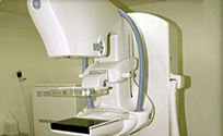
Digital 3D Tomosynthesis Mammography
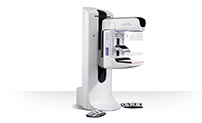
Mammography, which is admitted as a domestic new technology, with the world first FDA approval of Hologic Inc.
It is a technology to film the breast in 5 seconds in 15 degrees and realize the image in three dimensions. It is a cutting edge breast diagnosis system which increased the accuracy of diagnosis of breast cancer.
- Tomography image filming in 1mm distances to improve accuracy
- Increase the detection rate of breast cancer, and reduce the re-examination rate to reduce time and cost
Surgery robot operator 'Da Vinci-Xi (da Vinci-Xi)'
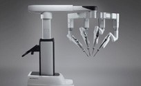
The latest model with supplementing the disadvantages of previous surgery robot model and upgrading the advantages, so safety and convenience is increased.
- As the robot’s arm got longer and thinner, robots are able to handle more deep and various degrees of operation
- Optical 3D solid high-resolution image provided.
- Reduction of the preparation process and anesthesia time during the surgery
- Fewer scars and quick recovery
In general, robot operation can treat diseases of a prostate, cervix, uterus, colon/rectal cancer, and thyroid gland with minimum cutting, so it is efficient in complex and difficult surgeries.
True BEAM
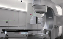
Truebeam, an extremely accurate and powerful cancer remover, can heal 2.5 mm-sized tumor without damaging the normal tissues and organs at all. By high-output of 2400MU/min, which is 4 times much higher than general equipment, reduces time and period of treatment.
Also, the medical staff can check the treatment status in real time and automatic recognition system of the physical movement is included, accuracy and safety of the treatment are increased.
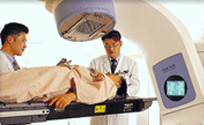
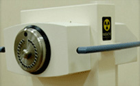
Radiotherapy device (21EX, Linear Accelerator)
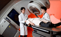
With a linear accelerator that can generate 6.15 MV X-Ray and 6,9,12,16,20MeV electron, it removes the tumor by exposing the radiation from the outside of the body, by using the latest treatment technologies like third-dimensional radiation and intensity-alterable radiation treatment.
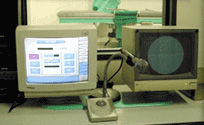
Simulator Device (Acuity, Simulator)
A simulator is a radiation penetration device that is made geometrically identical with the radiation treatment device. The equipment in our hospital is the first domestic simulator, and it included electronic video acquisition device so fast and accurate simulation is possible.
Proximity treatment device (GAMMAMED PlusⅡ)
This device has the advantage of protecting surrounding normal tissues efficiently by exposing a large amount of radiation directly to the part of the tumor as the treatment method of inserting the radiation source into the body.
SPECT gamma camera
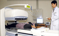
SPECT gamma camera combines the labeled compound with the radioactive isotopes, and collects the radiation from the radioactive isotopes by the gamma camera, and uses the computer to rebuild the organs in the human body in 3 dimensions, and visualizes the organ’s change and physiological function, so it has prominent effect on considering the diagnosis and treatment deep part’s disease.
Especially, it has prominent diagnosis effect on the metabolic disorder of organs, cardiovascular disorders, cerebrovascular disease, and it can find the reason of the epileptic seizure to cure it by surgery, and it is also used in early cancer diagnosis and observation.
PET - CT
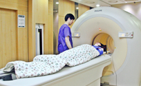
PET – CT a cutting-edge diagnosis equipment made of a combination of Positron Emission Tomography(PET) and Computed Tomography(CT) that can diagnose cancer in the whole body with just one examination, and find out the location of it. It diagnoses the metabolic load, activity of cancer so even small cancers with high malignity can be found accurately, so it is used in tumor diagnosis like early diagnosis of cancer, treatment response evaluation, process and invasion range, tumor’s remote transfer, or neuro-psychosis like Alzheimer's disease(Dementia), epilepsy, cerebral palsy, mental illnesses (depression, obsession, and schizophrenia), stroke, and cerebrovascular.
CT (COMPUTED TOMOGRAPHY)
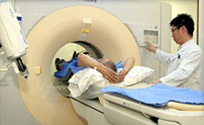
CT is an imaging technology that collects the X-rays exposed from various directions to penetrate the goal part of body with a detector, and analyzes the X-ray absorption difference with a computer by using math methods. Compared to the X-ray pictures in the past, it has prominent resolution and contract in classifying the blood and cerebrospinal fluid, white matter, tumors, tumor, and it can express the absorption difference of the subtle parts, so it is playing an important role in image diagnostic method.
The basic principle is the X-ray tube spins around the cross section of the body and exposes the X-ray beam, and the detector collects the intensity, and the computer calculates the absorption intensity from each part by the data to rebuild a video and display it on monitor. MDCT in Ulsan University hospital has 16 multiple detectors so that it can image a wide range of examination area quickly in a light thickness compared the existing CTs which has only one detector.
After collecting the image, by follow-up management process using an upgraded computer, we are able to provide medical service of good quality by CT blood vessel image (CT-ANGIO) D and 3-dimensional video with more accuracy and daily availability. It can image the moving organs like heart, and it can create 3 and 4-dimensional video for diagnosing the disease solidly and accurately.
Digital angiogram
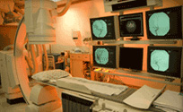
An angiogram is imaging the blood vessel by inserting fine tube-like noodles called catheter and using contrast medium and angiogram device. This is a basic image technology used in all vessel intervention procedure
Intervention procedure
Various intervention procedures are processed in radiology department.
- 1. Angiogram
- 2. Percutaneous angioplasty of peripheral vascular disease
- 3. Clotbuster treatment of deep vein thrombosis
- 4. Nonoperative treatment of aortism: Artificial blood vessel stent
- 5. Nonoperative treatment of uterine myoma: Pelvic venography embolization
- 6. Nonoperative treatment of chronic pelvic pain: Pelvic venography embolization
- 7. Nonoperative treatment of liver cancer
- 8. Nonoperative treatment of diseases of biliary tract
- 9. Nonoperative treatment of urosis
- 10. Nonoperative treatment of gastrointestinal tract
- 11. Various biopsies
- 12. Nonoperative treatment of cerebrovascular disease
Angiogram device of Siemens company, Germany in Ulsan university hospital is a cutting edge treatment and diagnosis device with the prominent definition. It reduces the procedure time and uses time efficiently because it can obtain an image without distortion due to the lesion’s curb, so it helps to provide the best medical service.
MRI (MAGNETIC RESONANCE IMAGING)
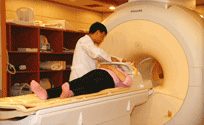
MRI, MAGNETIC RESONANCE IMAGING examines the patient in a device composed of strong magnet and exposes Radio Frequency Pulse using the magnetic field and analyzes the signal from the Hydrogen nucleus in the body to make the video by calculating the resonance difference of each tissue and organ.
In short, this is a diagnostic method rebuilding the measured magnetic characteristic of the materials composing the body by the computer and visualizing it. Existing radiation image for diagnosis uses the radiation, but it is the biggest difference that MRI uses a magnet.
MRI makes us obtain videos from various surfaces, and understand the materials of the lesion with prominent contrast, so it is the best video examination method in many diseases including Cranial nerve and muscle and bone system. Achieva MR 3.0T of Philips in Ulsan university hospital is the latest model in clinic, and it reduces the examination time so to reduce the pressure of Emergency patient, claustrophobia patients, or patients without self-control.)
It reduced the risk of side effects by minimizing the quantity of contrast medium for the patient.
Compared to existing devices, the signal intensity and definition are double higher, so it diagnosed the fine structure accurately.
Using the special video method, you can diagnosis the anatomical construction change of cerebrosis like Epilepsy, dementia.
Provides prominent information of early diagnosis and treatment by measuring the metabolite change of tumors and metabolic disease.
Ultrasonic wave impact crusher
COMPACT-SIGMA, Ultrasonic wave impact crusher in our hospital, is the latest model of Dornier, Germany, which invented the external Ultrasonic wave impact crusher for the first time in 1980, and the first in market share in the world, and it is praised for its name value.
It is similar with the ideal shockwave which has the short rise time and doesn’t have sound pressure, and it’s safe and efficient shock wave using the Electromagnetic method. By the best crushing technology, it takes short time to do the procedure. Hospitalization is not necessary since it is not a surgery.
There is no pain in the procedure and anesthesia is not necessary, so there are no physical risk, complications, and side effects due to surgery or anesthesia. By the best crushing effect, the disease is cured perfectly in a short time, and re-treatment rate is low, so the reliability of the treatment is increased. It is very safe for the patient because it does not affect other organs in the body, and it does not interrupt the daily life.
Digital mammography device
Mammography is the most basic examination in the breast cancer screening.
Digital Mammography device in our hospital obtains image with higher definition compared to the existing analog Mammography device for accurate diagnosis, and decoding is possible in a short time due to digital video treatment.
In mammography, many women felt uncomfortable before. After the introduction of digital mammography device, it reduces the pressure and the radiation exposure.
Also, it is an innovative device that can immediately operate stereotaxic mammotome tissue biopsy for micro-calcification, which can often be seen in early breast cancer.